Research
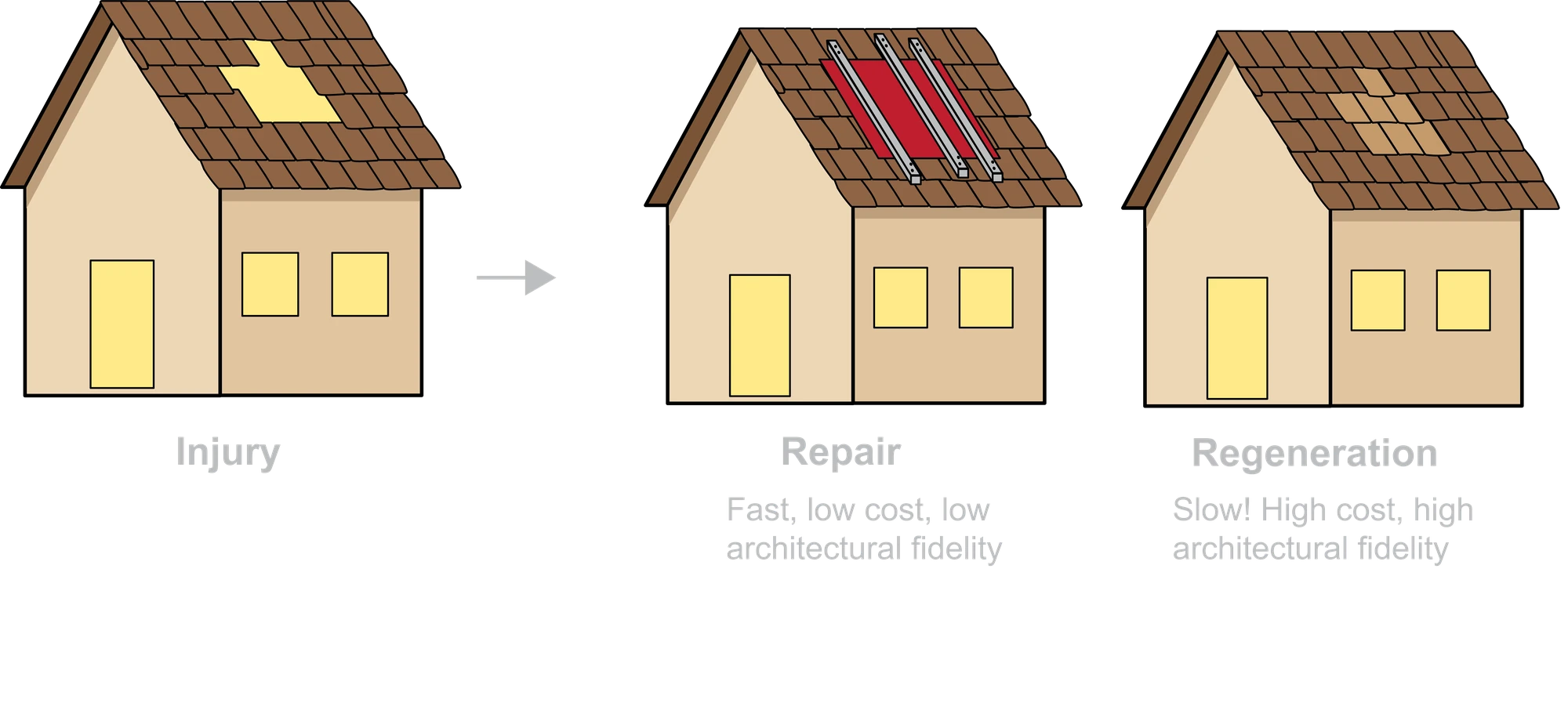
Life is full of risks. From microbes to major accidents, injury is a challenge every organism faces, with profound biological consequences. Injury and repair are of ancient origin, conserved from sea urchins to mammals. In an ideal world, the immune system detects damage and triggers a regenerative response that restores both tissue function and architecture ad integrum. In most mammals, repair is fast but imperfect — it leaves a scar.
We study what happens at the interface of immunity, tissue repair, and the body cavities. Our work has clinical roots — many of the questions we ask come from problems we encountered in the operating room — but our methods are squarely in mechanistic basic science.
Themes
Cavity Macrophage Aggregation
Within minutes of injury, free-floating GATA6⁺ macrophages vanish from the peritoneal fluid and pile onto the wound — the macrophage disappearance reaction. We showed this aggregation is driven by the scavenger receptors MARCO and MSR1, depends on calcium, and is conserved all the way to sea urchin coelomocytes. We now ask which ligand engages these receptors, why the cells don’t aggregate spontaneously, and how their post-injury fate shapes repair versus scarring.
Mesothelial to Mesenchymal Transition
After surgery, healthy mesothelial cells shed their epithelial identity, turn migratory, and reprogram into the myofibroblasts that lay down scar. We’ve mapped part of this transition and shown it depends on cues from the peritoneal microenvironment, including microbial contamination and EGFR signaling. We are now dissecting how mesothelial cells are recruited, how immune crosstalk tips the balance between physiological repair and pathological scarring, and building transplantation systems for genetically engineered mesothelial cells.
Foreign Body Reaction and Implant Lifetime
Surgical injury rarely comes alone — it usually arrives with foreign material: sutures, meshes, implants. The combination of tissue damage and biomaterial fundamentally changes how repair proceeds. Working with bioengineers, we study how implant surface properties shape the local immune response, aiming to design materials that integrate rather than scar.
Translational Studies
We connect mechanistic discovery to human biology through prospective patient cohorts (with surgical partners at Inselspital Bern) and a bio-banked pipeline of fresh and frozen human peritoneal cells. A particular interest is the translational gap between preclinical and human peritoneal macrophages. We have been involved in start-ups and consult for established pharmaceutical companies.
Methods
Intravital Microscopy
The signature method of the lab. We image immune cells and mesothelial dynamics in real time, including through the intact abdominal wall — an approach originally developed in the Kubes lab and now extended in Zürich. Our setup is a Leica Stellaris DIVE multiphoton system with a 12 kHz resonant scanner, hybrid detectors, and full FLIM (fluorescence lifetime imaging), letting us read out cellular metabolism and signaling states alongside behavior.
Quantitative Image Analysis
Beautiful imaging is only the starting point. We complement our microscopy with custom in-house pipelines for next-generation cell segmentation, classification, and tracking, run on ETH’s high-performance computing infrastructure. These tools turn 4D intravital movies into reproducible, cell-by-cell quantification — bridging visually striking microscopy with the statistically tractable outcomes that mechanistic conclusions require.
Single-Cell and Spatial Transcriptomics
We map cell states across preclinical and human peritoneal tissues using scRNA-seq, regulon inference (pySCENIC), and spatial transcriptomics — identifying candidate regulators of mesothelial and macrophage behavior.
Models of Peritoneal Injury and Repair
We developed and published two complementary in vivo models of peritoneal repair: the focal peritoneal injury (FPI) model, which follows how a wound resolves, and the peritoneal button (PB) model, which captures how adhesions form. Combined with transgenic, reporter, and lineage-tracing approaches, they let us interrogate the specific cellular contributions to healing versus scarring.
Human Peritoneal Sampling
Through clinical collaborations we run a robust pipeline for fresh human peritoneal fluid and tissue, enabling direct cross-species validation of our preclinical findings.
Research in Animals
Our research relies in part on animal experimentation, and we approach this responsibility with compassion and rigor. Every experiment we conduct is approved by the cantonal veterinary office after careful review by an ethics commission. This ensures that our work adheres to the 3R principles: wherever we can, we replace animal experiments, reduce the number of animals used, and refine our experimental approach — so that animals are involved only when no alternative can answer the scientific question, in the smallest numbers necessary, and under conditions that minimise distress.
We are convinced that responsible animal research remains essential. Much of what we understand about tissue repair, immunity, and disease — and many of the therapies that have followed — would not exist without it. For this reason, Joel Zindel is a member of Verein Forschung für Leben, a Swiss association of researchers — including colleagues from ETH Zürich — who advocate for the importance of biomedical research and for open, honest dialogue with the public.
Funding
We are grateful for the support of:
- Swiss National Science Foundation — Starting Grant
- Swiss National Science Foundation — Project Grant
- Swiss National Science Foundation — Weave
- Helmut Horten Foundation
- Geistlich Stucki Foundation
- ETH Foundation